 Self-cycleTM IVF


-
-
-
 Infertility Counselling
-
 Female Infertility Treatment
-
 Andrology Treatment
-
 Fertility Enhancing Surgeries - Female
-
 Fertility Enhancing Surgeries - Male
-
 Endoscopy Treatment
-
 IUI Treatment
-
 IVF Treatment
-
 ICSI Treatment
-
 Advance IVF Solutions
-
 Embryology
-
 Vitrification Egg, Embryo, Sperm Freezing
-
 Preimplantation Genetic Testing (PGT)
-
 Donation Program Embryo / Egg / Sperm
-
 Self-cycleTM IVF
-
-
-
-
-
Low Amniotic Fluid Pregnancy: Causes, Symptoms, and How to Increase Amniotic Fluid Safely
Low amniotic fluid in pregnancy, known medically as oligohydramnios, means the fluid around your baby has measured below the expected range for your stage. It is usually picked up on a routine ultrasound. Most cases produce no symptoms that you would notice yourself.
Learning that your fluid is low between scans is unsettling. The right response is prompt clinical assessment rather than alarm. Many mild cases are then monitored safely through the rest of pregnancy.
How much a low reading matters depends on the cause, the stage of pregnancy, and how low it is. Some cases need closer follow-up or a planned delivery. The interpretation belongs with your obstetric team, not with a single number.
The same fluid reading can mean different things at 28 weeks and at 40 weeks.
What Is Low Amniotic Fluid (Oligohydramnios)?
Amniotic fluid is the fluid that surrounds and protects your baby in the womb. It cushions the baby and the umbilical cord and helps guard against infection. The fluid also gives the developing lungs and limbs room to grow and move.
From the middle of pregnancy onward, most amniotic fluid is your baby's urine. This is why conditions affecting the fetal kidneys or urine flow can lower the volume. Before mid-pregnancy, the fluid comes mainly from the placenta and fetal membranes instead.
Oligohydramnios is diagnosed on ultrasound using one of two standard measurements. The amniotic fluid index adds the deepest fluid pocket in each of four sections of the uterus. The single deepest pocket instead measures the one deepest pool of fluid.
| Measurement | Oligohydramnios threshold | Typical normal range |
|---|---|---|
| Amniotic fluid index (AFI) | 5 cm or less (some centres use below 5) | greater than 5 up to about 24 cm |
| Single deepest pocket (SDP) | less than 2 cm | about 2 to 8 cm |
A low reading is a signal for closer assessment, not an automatic sign of harm to the baby. Its meaning depends on why the fluid is low and when in pregnancy it appears. A borderline result is often rechecked within a few days before any decision is made.
Amniotic fluid volume is a measurement taken during an established pregnancy. It sits within routine antenatal monitoring rather than pre-conception care. General fertility education covers the earlier assessments that precede conception, which are a separate topic.
Causes of Oligohydramnios: Why Amniotic Fluid Gets Low
The causes of low amniotic fluid in pregnancy usually fall into three groups: maternal, placental, and fetal. Maternal causes are the most often reversible, while fetal causes rarely are. Identifying the cause is what shapes the next steps in care.
Placental and Maternal Causes
On the maternal and placental side, reduced blood flow through the placenta limits the fluid the baby can produce. Common contributors include high blood pressure and pre-eclampsia, maternal dehydration, and poorly controlled diabetes. Certain medicines, such as some blood-pressure drugs and anti-inflammatory medications, can also lower the fluid.
Several of these factors are potentially correctable, particularly when found in the third trimester. Treating the underlying maternal condition can help stabilise the fluid. Rehydration, blood-pressure control, or reviewing a contributing medicine can each raise the fluid measurably.
Foetal and Structural Causes
On the baby's side, problems with the kidneys or urinary tract cut urine output. Urine is the main source of amniotic fluid from mid-pregnancy onward. A minority of these cases involve congenital or genetic conditions affecting those organs.
Preimplantation genetic testing can screen embryos for a known heritable condition before transfer. It is relevant only when a specific inherited condition runs in the family. The test screens embryos rather than treating low fluid in an established pregnancy.
A separate cause is the waters breaking early, called preterm prelabour rupture of membranes (PPROM). Its typical sign is a sudden gush or a steady trickle of clear fluid from the vagina. This needs a same-day medical review, because it changes how the pregnancy is managed.
PPROM is not the same as the usual increase in vaginal discharge late in pregnancy. Any leaking of clear fluid should be checked promptly. A simple speculum exam or a bedside test can confirm whether the membranes have ruptured.
Amniotic Fluid Low Symptoms: Signs to Watch For
Low amniotic fluid often causes no symptoms at all, and many cases are found only on a scan. A few changes can be noticed at home, though none confirm low fluid alone. They overlap with the ordinary sensations of late pregnancy, so context matters.
The possible signs to watch for include the following:
- A feeling that the baby is moving less than usual
- A bump that measures smaller than expected for your dates
- Any leaking of fluid, which always needs prompt checking
When Symptoms Need Immediate Attention
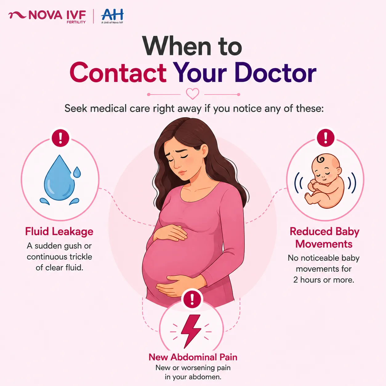
Some signs need same-day attention because they can reflect reduced oxygen supply to the baby. Contact your maternity unit straight away if you notice the signs below. Waiting for the next routine appointment is not advisable in these situations.
- A sudden gush or a continuous trickle of clear fluid
- No noticeable baby movements for two hours or more
- New or worsening abdominal pain
At the unit, the team monitors the baby's heartbeat with a CTG trace. An ultrasound then measures the fluid and reviews the baby's growth. Together these checks show whether the baby is currently coping well.
Complications of Low Amniotic Fluid in Pregnancy
When fluid stays low, the possible complications depend heavily on the stage of pregnancy. Earlier in pregnancy the concerns are greater, because fluid is needed for the lungs and limbs to develop. Later on, attention shifts to the umbilical cord and the baby's growth.
Reported associations from clinical studies include the following:
- Compression of the umbilical cord, which can reduce the baby's oxygen supply
- Slower growth, described clinically as fetal growth restriction
- Underdeveloped lungs when fluid is very low early in pregnancy
- A higher chance of induced labour or caesarean birth
A 2025 case-control study of idiopathic oligohydramnios in Eastern India, published in the Nigerian Medical Journal by Dutta and colleagues, compared low-fluid pregnancies with matched controls. Each arm held 50 pregnancies at 34 weeks or later. The low-fluid group had higher rates of caesarean birth, small-for-gestational-age babies, and NICU admissions.
Importantly, that study recorded no perinatal deaths in either group. The authors attributed this to close monitoring and timely delivery. Its findings apply to near-term, otherwise unexplained cases, not to every pregnancy.
Because the stage of pregnancy matters, accurate dating supports the interpretation of a low reading. An IVF due date calculator can help confirm the timeline of an IVF pregnancy. A confirmed date also guides when a planned delivery would be safe if one is needed.
How to Increase Amniotic Fluid During Pregnancy
Approaches to raising low amniotic fluid begin with simple measures and escalate only when the situation calls for it. The right choice turns on why the fluid is low and how far along you are. None of these steps replaces the assessment that finds the underlying reason.
Hydration and Rest: The First Steps
For some women, drinking more water is a reasonable first step, and there is evidence behind it. A 2002 Cochrane review by Hofmeyr, Gulmezoglu and Novikova found that maternal hydration raised the measured amniotic fluid index. The review pooled four small trials involving 122 women in total.
The pooled trials were small, so the certainty behind this benefit remains low. Those trials showed more fluid on the scan, but did not assess whether hydration improves the baby's outcomes. So hydration may support care, but it does not replace finding and treating the cause.
Left-side rest and adequate hydration are supportive measures only. They are not a treatment for oligohydramnios and not a substitute for specialist evaluation. Any low reading still needs an obstetric assessment to find why the fluid is low.
Medical Treatments for Oligohydramnios
When the cause or severity calls for more, treatment is tailored to the individual situation. Options can include intravenous fluids in hospital for significant dehydration. Another is amnioinfusion, where sterile fluid is placed into the amniotic sac during labour.
Near term, the obstetric team may recommend a planned delivery when that is the safer option. Gestational age, the underlying cause, and the baby's wellbeing all steer that decision. Very low fluid at or beyond 36 to 37 weeks often tips that balance toward delivery.
This monitoring applies whether you conceived naturally or through IVF. Pregnancies achieved with advanced IVF solutions are followed just as closely. Your fertility and obstetric teams coordinate the plan.
Frequently Asked Questions
What are the main causes of low amniotic fluid during pregnancy?
The causes fall into three groups: maternal and placental factors such as high blood pressure, pre-eclampsia and dehydration; fetal kidney or urinary problems; and the waters breaking early. In some pregnancies, no clear cause is found on assessment.
What are the warning signs of low amniotic fluid?
Often there are none, and low fluid is found on a scan. When signs appear, they may include reduced baby movements or a bump measuring small. A gush or trickle of fluid, absent movements, or abdominal pain need same-day review.
How can I increase amniotic fluid naturally?
Good hydration is the main step, and a 2002 Cochrane review linked it to a higher measured fluid index. Resting on your left side may also help. These support the readings but do not replace finding the cause or specialist assessment.
Is low amniotic fluid dangerous for the baby?
It depends on the cause, the stage, and how low the fluid is. Very low fluid early in pregnancy carries more risk, while many mild or near-term cases do well with monitoring. It needs assessment rather than assumption.