 Self-cycleTM IVF


-
-
-
 Infertility Counselling
-
 Female Infertility Treatment
-
 Andrology Treatment
-
 Fertility Enhancing Surgeries - Female
-
 Fertility Enhancing Surgeries - Male
-
 Endoscopy Treatment
-
 IUI Treatment
-
 IVF Treatment
-
 ICSI Treatment
-
 Advance IVF Solutions
-
 Embryology
-
 Vitrification Egg, Embryo, Sperm Freezing
-
 Preimplantation Genetic Testing (PGT)
-
 Donation Program Embryo / Egg / Sperm
-
 Self-cycleTM IVF
-
-
-
-
-
Ovarian Follicles: What They Are, How They're Counted, and What It Means for Your Fertility
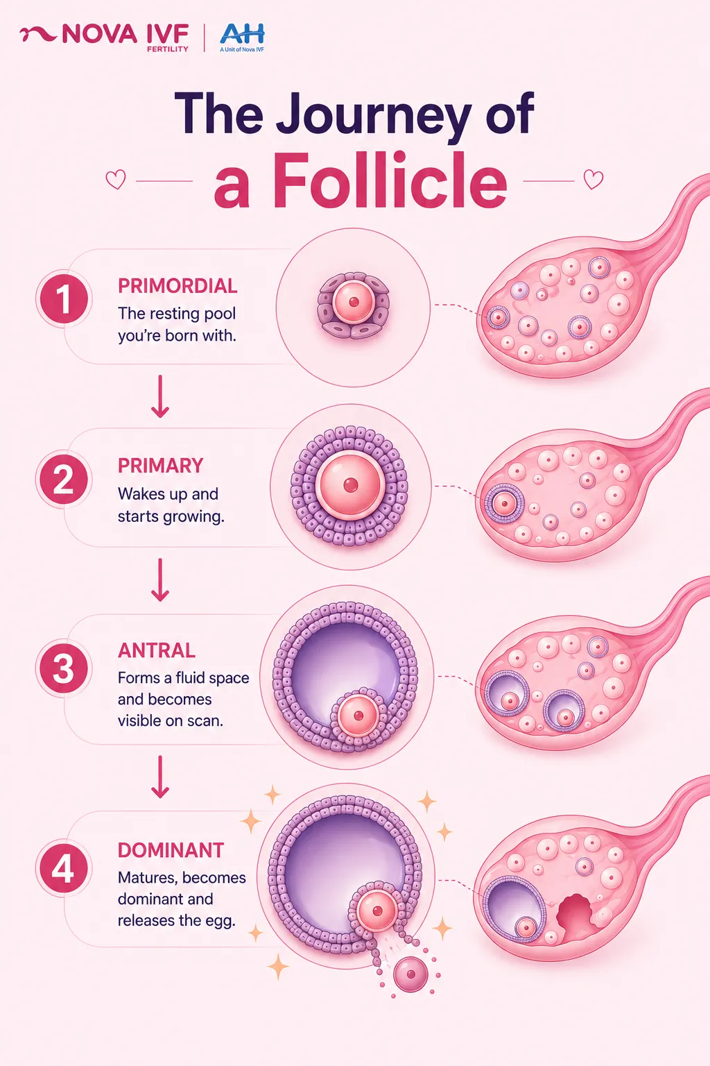
Ovarian follicles are small, fluid-filled sacs inside the ovaries, and each one holds a single immature egg. The cells lining each follicle nourish the egg and control when it matures. They also release hormones such as estrogen that help drive the menstrual cycle.
The follicle count is a useful marker of how many eggs a woman has left. A count can look alarming on a scan report, yet it shows only that month's resting follicles. What a result means depends on age, hormones, and cycle history, not the number alone.
This content is for informational purposes and does not substitute medical advice. Individual clinical situations vary, and treatment decisions should be made in consultation with a qualified reproductive medicine specialist. A follicle count on its own is not a diagnosis.
What Are Ovarian Follicles and How Do They Work?
A follicle is the egg's container, not the egg itself. It holds one egg but does not guarantee that egg will mature into a usable one. On a scan report, a follicle is counted whether or not the egg inside will ever ovulate.
According to reproductive-biology research first quantified by Baker in 1963, a woman is born with roughly 1 to 2 million follicles. Current ovarian-reserve reviews still report this range. The supply then falls steadily with age, which is why follicle counts sit at the centre of most fertility checks.
Each month, the brain releases follicle-stimulating hormone, known as FSH, which prompts a batch of follicles to grow. Usually only the one most ready to respond becomes dominant, while the others stop and fade. This monthly selection is normal and repeats from puberty until menopause.
Stages of Follicle Development: From Primordial to Dominant
Follicles move through clear stages before one of them releases an egg:
- Primordial follicles are the dormant, resting pool present from birth.
- Primary and secondary follicles wake up, begin to grow, and start producing hormones.
- Antral follicles develop a small fluid space and become visible on a scan.
- Usually one follicle pulls ahead of the rest and is called the dominant, or Graafian, follicle.
- The dominant follicle releases its egg at ovulation, which is why the timing of ovulation sets the fertile window.
Antral Follicle Count (AFC): What It Is and Why It Matters
The antral follicle count, or AFC, is the number of resting follicles on a scan, each roughly 2 to 9 millimetres wide. A follicle count ultrasound is usually done on day 2 to 5 of the cycle. It uses a transvaginal probe to view both ovaries.
The AFC is one of the measures recorded during a female fertility assessment. It helps estimate ovarian reserve, the pool of eggs a woman has left. Doctors read it alongside other markers rather than on its own.
| Total AFC (both ovaries) | What it suggests |
|---|---|
| Below 6 to 7 | Low ovarian reserve, fewer eggs available |
| 8 to 15 | Average to good reserve |
| Above 20 to 30 | High count, sometimes linked to polycystic ovary syndrome (PCOS) |
These bands are approximate and depend on population, scanner, and clinic. A high count points to PCOS mainly when it is high for a woman's age. Doctors read the number alongside age, AMH, and cycle history.
A low count does not mean a woman cannot conceive, since age and egg quality also matter. Where reserve is low, some women consider egg freezing sooner rather than later. The count is usually read alongside the AMH blood test, which measures a hormone linked to egg supply.
Average Antral Follicle Count by Age: What's Normal?
The average antral follicle count by age falls gradually as the egg supply declines, a pattern shown in age-specific nomograms from Indian women. Under 35, a woman often has around 15 to 30 follicles in total. Between 35 and 40 this typically eases to about 10 to 15.
Counts then tend to fall to roughly 5 to 10 between 40 and 44, and often below 5 after 44. These figures are approximate ranges, not fixed cut-offs, and they vary by population, clinic, and scanner. A specialist reads a result alongside age, AMH, and history before drawing any conclusion.
Both Ovaries Show Multiple Small Follicles: What Does It Mean?
When both ovaries show multiple small follicles, a scan often describes a string of pearls around each ovary. This means many immature follicles are visible at once, a picture called polycystic ovarian morphology, or PCOM. Many women with this finding have regular cycles, normal hormones, and no health problems.
It Does Not Mean a High Egg Count
The follicles are real, but they begin growing and then stall before one matures and ovulates. Only one usually completes growth and releases an egg each cycle, so a crowded scan does not mean many ready eggs. A visible count reflects follicle pattern, not the number of mature eggs.
When This Points to PCOS
Polycystic morphology on its own does not confirm PCOS, and the two are often confused. A diagnosis of polycystic ovary syndrome needs two of three features. These are irregular cycles, raised male hormones, and the polycystic follicle pattern.
Older ultrasound criteria flagged 12 follicles per ovary. The 2023 International Evidence-based Guideline for the Assessment and Management of PCOS raised this to 20 follicles per ovary with modern high-resolution scanners. The scan alone still cannot make the diagnosis.
The next step is a review of cycle regularity alongside a blood test for testosterone and other androgens. That combination separates a benign follicle pattern from PCOS that needs care. Treating the scan label alone, without cycle and hormone data, drives the over-diagnosis that specialists warn against.
Follicle Count in IVF: What to Expect at Nova IVF
In IVF, the antral follicle count signals how the ovaries may respond to stimulation. A higher count tends to be associated with more eggs at collection, though it is only part of the picture. Nova IVF checks the count within a personalised workup before any plan is set.
- A count in the average range is generally associated with a typical response, though dose and protocol are still individualised.
- When the count is low, fewer eggs may be retrieved, so the dose or protocol is adjusted.
- Very high counts, often linked with PCOS, call for careful lower-dose stimulation to reduce the risk of OHSS, or ovarian hyperstimulation syndrome, a serious over-response.
- During an IVF cycle, repeat scans track follicle growth so timing and dose stay appropriate.
- The count is most informative when read with the AMH blood test and a woman's age.
Frequently Asked Questions
Is it normal to see follicles on an ultrasound?
Yes, seeing follicles on a scan is completely normal and expected. Several small antral follicles appear in each ovary at the start of every cycle. Their number helps estimate ovarian reserve, not whether something is wrong.
What is the difference between a follicle and an egg?
The follicle is the fluid-filled sac, and the egg is the cell held within it. A single follicle contains one egg, though it may grow and still fail to release a mature one. Counts therefore reflect egg supply, not egg quality.
Does four follicles mean four eggs?
Not reliably, because a follicle contains an egg but does not guarantee a mature, usable one. Some follicles develop without releasing a healthy egg. Four follicles is therefore a positive indication rather than four confirmed eggs.
How many follicles are normal at age 37?
Around 35 to 40, a total of roughly 10 to 15 antral follicles is typical. A figure a little above or below this rarely means high or low fertility on its own. Age and the AMH blood test give the fuller answer.
Can the antral follicle count be increased?
The count mostly reflects the eggs you already have, so it does not rise in a lasting way. Healthy habits support egg quality but do not rebuild the resting pool. This is why testing early, rather than waiting, is often useful.
Does a high follicle count always mean PCOS?
No, a high count does not always mean PCOS, and many women with many follicles are healthy. PCOS is confirmed only when the follicle pattern appears with irregular cycles or raised male hormones. The count alone is one clue, not a diagnosis.
How are multiple small follicles diagnosed?
Multiple small follicles are first spotted on a routine pelvic or fertility scan. The radiologist counts the follicles in each ovary and notes their size and pattern. A specialist then combines that scan with your cycle history and hormone tests.
Related Fertility Blogs