 Self-cycleTM IVF


-
-
-
 Infertility Counselling
-
 Female Infertility Treatment
-
 Andrology Treatment
-
 Fertility Enhancing Surgeries - Female
-
 Fertility Enhancing Surgeries - Male
-
 Endoscopy Treatment
-
 IUI Treatment
-
 IVF Treatment
-
 ICSI Treatment
-
 Advance IVF Solutions
-
 Embryology
-
 Vitrification Egg, Embryo, Sperm Freezing
-
 Preimplantation Genetic Testing (PGT)
-
 Donation Program Embryo / Egg / Sperm
-
 Self-cycleTM IVF
-
-
-
-
-
Fetal Development Week by Week: A Complete Guide from Week 1 to Week 40
Finding out you are pregnant begins a life-changing journey. You will likely feel a mix of excitement, wonder, and curiosity as you imagine how your baby grows and changes inside you each day. Every tiny flutter, new symptom, and prenatal visit brings you one step closer to holding your baby.
Tracking fetal development week by week turns your curiosity into clear understanding. From early cellular growth in the first month to the final preparations for birth, your baby reaches incredible milestones in organ development, movement, and size.
This guide walks you through fetal development across all three trimesters. You will discover key milestones from Week 1 to Week 40 and learn how regular prenatal care protects you and your baby throughout your pregnancy.
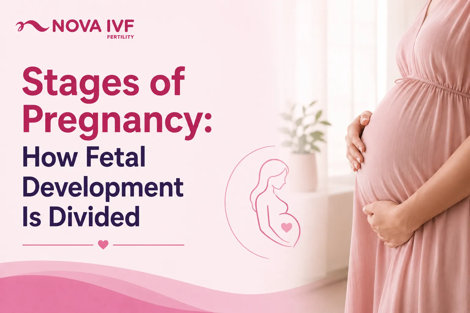
Stages of Pregnancy: How Fetal Development Is Divided
A full-term human pregnancy lasts approximately 40 weeks (280 days), calculated from the first day of your last menstrual period. Healthcare providers divide these 40 weeks into three distinct trimesters:
- First Trimester: Week 1 to Week 12
- Second Trimester: Week 13 to Week 27
- Third Trimester: Week 28 to Week 40 (or delivery)
Understanding these 3 stages gives you a clear plan for what to expect during your prenatal visits, scan appointments, and daily physical changes. For parents who conceived through IVF, an IVF due date calculator can help estimate the pregnancy timeline and expected due date.
First Trimester of Fetal Development (Weeks 1–12)
The first trimester spans gestational Weeks 1 to 12 and includes rapid embryonic and fetal development. Key developmental milestones during the first trimester include:
- Neural Tube Formation:
By week 4 to week 5 of pregnancy, the neural tube closes. The neural tube forms the foundations of the baby's brain and spinal cord.
- Early Cardiac Activity:
Around week 6 of pregnancy, early cardiac cells begin rhythmic contractions, which can often be detected via transvaginal ultrasound.
- Embryo to Fetus Transition:
During the early stages of development, the developing baby is medically termed an embryo. From approximately the ninth week after conception, it is classified as a fetus as development shifts toward continued growth and maturation.
- Organ Development:
By the end of the first trimester, the basic structures of major organ systems, including the brain, heart, kidneys, liver, and limbs, have formed and continue to mature throughout pregnancy.
Because early pregnancy involves rapid cellular and developmental changes, the risk of pregnancy loss is higher during this period. A scientific review published in Translational Pediatrics reports that approximately 10% to 15% of clinically recognized pregnancies end in spontaneous loss, with many early losses associated with chromosomal abnormalities. As pregnancy progresses beyond the first trimester, the statistical risk of miscarriage generally decreases.
Unexpected or persistent bleeding during pregnancy should be discussed promptly with a healthcare provider. For individuals with a history of repeated pregnancy loss, discussing recurrent miscarriage treatment options with a fertility specialist may help identify and manage potential contributing factors.
Second Trimester of Fetal Development (Weeks 13–27)
The second trimester of fetal development spans weeks 13 to 27 and involves rapid physical growth, improved muscle coordination, and system maturation. Early gestational symptoms like morning sickness and severe fatigue often ease during this period, making it a comfortable phase for many expectant mothers.
Key physical developments during the second trimester include:
- Fetal Movement (Quickening):
Mothers typically feel the first subtle fetal movements, known as quickening, between weeks 18 and 22. Initial movements feel like gentle flutters and grow stronger as fetal muscle mass increases.
- The Mid-Pregnancy Anatomy Scan:
A detailed ultrasound is performed between weeks 18 and 22 and evaluates physical development. The scan checks the structure of the heart, brain, spine, kidneys, and placenta, while also allowing parents to learn the baby's biological sex if desired.
- Hearing and Sensory Growth:
The auditory system continues to mature during the second trimester, and the fetus becomes increasingly responsive to sounds from inside and outside the womb, including the mother's voice and heartbeat.
During the second trimester, fetal facial features become distinct, skin remains protected by a fine coating called lanugo, and finger pads form unique fingerprints by the end of this trimester.
Third Trimester of Fetal Development (Weeks 28–40)
The third trimester of fetal development spans from week 28 until birth and focuses on rapid weight gain, lung maturation, and preparing for life outside the womb. During these final months, the fetus gains roughly 200 grams per week, accumulating fat layers under the skin to help with thermal regulation after birth.
Key fetal developments occurring in the third trimester include:
- Lung Maturation:
The fetal lungs begin producing pulmonary surfactant, a protein-lipid compound that keeps the air sacs (alveoli) open.
- Cephalic Positioning:
Between weeks 32 and 36, many babies settle into a head-down (Cephalic) position, which is the most common position for vaginal birth.
- Neurological Growth:
Brain tissue expands rapidly, creating complex neural networks. The fetus establishes recognizable sleep-wake cycles and responds actively to light and acoustic stimuli.
A pregnancy reaches full-term status at 39 weeks. Although babies born from 37 weeks onward are considered term, brain, lung, and other organ development continues during the final weeks of pregnancy.
Fetal Development Stages: Week by Week Milestones to Watch For
Tracking fetal development week by week helps healthcare providers and expectant parents understand how development progresses throughout pregnancy. The following table provides an overview of major developmental milestones from the first weeks of pregnancy through birth:
| Gestational Timing | Developmental Milestone | What It Means |
|---|---|---|
| Weeks 1–2 | Pregnancy dating, ovulation and fertilisation | Pregnancy is dated from the first day of the last menstrual period. Ovulation and fertilisation generally occur around Week 2, although timing can vary. |
| Weeks 3–4 | Implantation and early embryonic development | The fertilised egg implants in the uterus, and early embryonic structures begin to develop. |
| Weeks 5–8 | Neural tube and early organ development | The neural tube develops into the brain and spinal cord, early cardiac activity begins, and the foundations of major organs start forming. |
| Weeks 9–12 | Embryo-to-fetus transition and organ development | The developing baby is classified as a fetus, while the basic structures of major organs, limbs, and facial features continue developing. |
| Weeks 13–17 | Rapid growth and movement | The fetus grows rapidly, develops stronger muscles, and begins making increasingly coordinated movements. |
| Weeks 18–22 | Fetal movement and anatomy assessment | Fetal movements may become noticeable, while the mid-pregnancy anatomy scan evaluates major organs, structures, and growth. |
| Weeks 23–27 | Sensory and organ maturation | The nervous system, lungs, and other organs continue developing as the fetus gains weight and responds increasingly to sounds. |
| Weeks 28–32 | Brain and lung development | Brain development accelerates, while the lungs continue maturing and the fetus gains body fat. |
| Weeks 33–36 | Weight gain and positioning | The fetus continues gaining weight and many babies settle into a head-down position as the body prepares for birth. |
| Weeks 37–40 | Final maturation and preparation for birth | The fetus continues gaining weight and undergoing final brain, lung, and other organ maturation before birth. |
Why Regular Prenatal Checkups Matter at Each Stage
Regular prenatal checkups help healthcare providers monitor fetal development week by week, assess maternal health, and identify potential concerns as pregnancy progresses. The type and frequency of assessments can vary based on the stage of pregnancy and individual health needs.
Key aspects of prenatal care across the three trimesters include:
First Trimester:
Early checkup appointments may include pregnancy confirmation, gestational age assessment, maternal health evaluation, and relevant blood tests. A dating ultrasound may also help establish the expected due date and assess early pregnancy development.
Second Trimester:
Prenatal visits continue to monitor maternal and fetal health, while the anatomy scan, usually performed around 18 to 22 weeks, evaluates the development of major fetal structures and the position of the placenta.
Third Trimester:
Checkups focus on fetal growth, position, maternal blood pressure, and overall fetal well-being. As delivery approaches, healthcare providers may also discuss birth planning and the care needed during the final weeks of pregnancy.
For those who conceived through IVF, preimplantation genetic screening may have been performed before embryo transfer to assess embryos for certain chromosomal abnormalities.
Every pregnancy follows its own course, so having appropriate guidance throughout each stage can help you understand the changes taking place and the care you may need. Nova IVF Fertility provides pregnancy and reproductive care with specialist guidance to help expectant parents with prenatal monitoring and make informed decisions throughout pregnancy.
FAQs - Frequently Asked Questions
In which week is the fetus fully developed?
Fetal development continues throughout pregnancy, and there is no single week when every organ suddenly becomes fully developed. A pregnancy is considered full term from 39 weeks 0 days through 40 weeks 6 days. By this stage, most major organ systems are sufficiently mature to support life outside the womb.
Which week is the hardest week in pregnancy?
The most challenging period varies for each individual. Many find weeks 6 through 10 difficult due to intense morning sickness, fatigue, and hormonal shifts. Others find weeks 36 through 40 challenging because of physical heaviness, pelvic pressure, and sleep disruptions.
How many weeks is full-term pregnancy?
A full-term pregnancy ranges from week 37 to week 40. Deliveries between 37 weeks 0 days and 38 weeks 6 days are classified as early term, while deliveries between 39 weeks 0 days and 40 weeks 6 days are classified as full term.
What are the most important weeks for fetal development?
Weeks 3 through 8 are critical for fetal development. During this embryonic stage, rapid cell division creates the neural tube, heart structure, and foundational organ systems, making the embryo sensitive to external environmental factors.