 Self-cycleTM IVF


-
-
-
 Infertility Counselling
-
 Female Infertility Treatment
-
 Andrology Treatment
-
 Fertility Enhancing Surgeries - Female
-
 Fertility Enhancing Surgeries - Male
-
 Endoscopy Treatment
-
 IUI Treatment
-
 IVF Treatment
-
 ICSI Treatment
-
 Advance IVF Solutions
-
 Embryology
-
 Vitrification Egg, Embryo, Sperm Freezing
-
 Preimplantation Genetic Testing (PGT)
-
 Donation Program Embryo / Egg / Sperm
-
 Self-cycleTM IVF
-
-
-
-
-
Gravid Uterus Explained: USG Report, Stages and Symptoms
If you have recently had an ultrasound and noticed the words “gravid uterus” on your scan report, you might wonder whether this finding is normal or if it points to a problem. Reading medical wording can feel overwhelming, especially when you are trying to understand how your pregnancy is progressing.
In almost every case, a gravid uterus simply means that your uterus is carrying a pregnancy. The term itself is not a warning sign, a diagnosis, or a complication. It is a standard medical term confirming a healthy pregnancy is underway.
This guide explains what a gravid uterus means, how the uterus changes during pregnancy, what you may see on a USG report, and when an ultrasound finding may need further medical review.
What Is a Gravid Uterus?
A gravid uterus is a uterus that currently contains a developing embryo or fetus. The word "gravid" originates from the Latin word gravidus, which translates to heavy, burdened, or pregnant. In clinical practice, obstetricians and sonographers use this term on routine antenatal reports to confirm that an intrauterine pregnancy is actively underway.
The uterus undergoes massive physical changes to accommodate a growing fetus. Before conception, a non-pregnant uterus is a small, hollow, pear-shaped organ located in the pelvis. It weighs approximately 50 to 70 grams and measures around 7.5 centimeters in length.
As pregnancy advances, the muscular walls of the uterus expand, stretch, and thin out. According to clinical studies, by the time a pregnancy reaches full term, the uterus grows into an organ weighing nearly 1,000 grams (1 kilogram) with a volume capacity thousands of times greater than its original state.
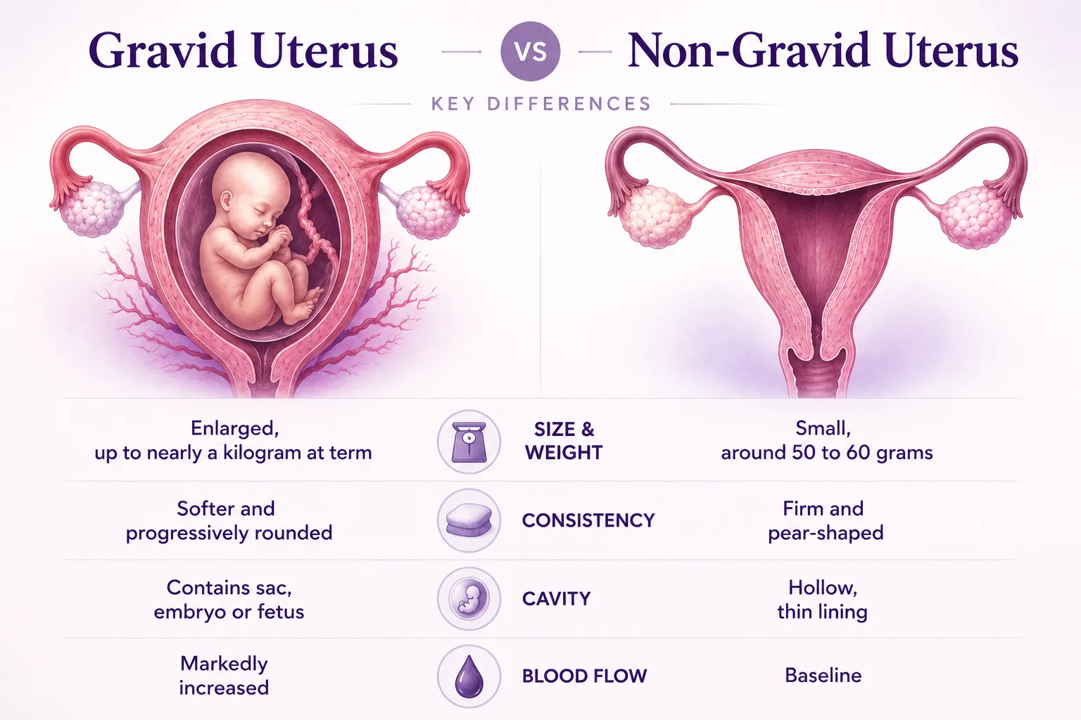
Gravid Uterus vs Non Gravid Uterus: Key Differences
Reports sometimes name a non-gravid uterus, meaning the resting, non-pregnant organ. Naming it tells the clinician the cavity holds no pregnancy. The same uterus becomes gravid once it adapts to carry one.
| Feature | Non-gravid uterus | Gravid uterus |
|---|---|---|
| Pregnancy Status | Absent | Present (Embryo/Fetus inside) |
| Size and weight | Small, around 50 to 60 grams | Enlarged, up to nearly a kilogram at term |
| Organ Consistency | Firm and pear-shaped | Softer and progressively rounded |
| Uterine Cavity | Hollow, thin lining | Contains sac, embryo or fetus |
| Blood flow | Standard baseline pelvic flow | Significantly increased circulation |
How a Gravid Uterus Changes Through the Trimesters
The gravid uterus grows in an almost predictable pattern across the three trimesters. In early pregnancy, it stays within the pelvis, so the change is felt more than seen. From the second trimester, it rises into the abdomen, and the bump becomes visible.
| Trimester | Uterus position | Typical landmark |
|---|---|---|
| First (weeks 1 to 12) | Stays within the pelvis | Rises to the pubic bone by about 12 weeks |
| Second (weeks 13 to 27) | Moves up into the abdomen | Reaches the navel by around 20 weeks |
| Third (weeks 28 to 40) | Fills much of the abdomen | Nears the ribs, then settles slightly before term |
Clinicians track this growth by measuring your fundal height: the distance from your pubic bone to the top of your uterus. This measurement offers a quick way to check your baby's growth between doctor visits. If a measurement falls outside the expected range, your doctor will order an ultrasound to take a closer look.
Common Symptoms as the Uterus Grows
The symptoms linked to a gravid uterus are the familiar signs of pregnancy itself. They appear as the organ enlarges and presses on nearby structures. Their intensity varies widely between women.
- A missed period followed by a positive pregnancy test
- Mild pelvic pressure or a sense of fullness low down
- More frequent urination as the uterus presses on the bladder
- A visible bump emerging through the second trimester
These changes are physiological rather than warning signs on their own. New or severe pain, vaginal bleeding, or a sudden change in symptoms is the exception that deserves prompt review. If in doubt, it is always reasonable to call your clinician.
How a Gravid Uterus Is Seen and Tracked on Ultrasound
Ultrasound is the main tool for confirming and following a pregnancy. Fertility ultrasound testing relies on the same imaging principles that later track a growing baby. A single scan can confirm the pregnancy's location, its stage, and the number of babies.
USG of Gravid Uterus: What the Ultrasound Shows
A USG of gravid uterus first confirms that the pregnancy sits inside the uterus, not in a fallopian tube. That finding rules out an ectopic pregnancy, which needs urgent care. The same scan identifies the gestational sac, the fetal pole, and later the heartbeat.
A gravid uterus ultrasound also estimates gestational age and checks how many babies are present. It notes the baby's position and where the placenta has settled. Each of these findings feeds directly into your antenatal plan.
Related Terms You May See on the Report
A report rarely stops at the gravid uterus, so other descriptive terms usually sit alongside it. Most are routine and simply record what the scan found. Each of the terms below points to a normal, expected feature of early pregnancy.
- Gestational sac: the fluid-filled space that holds the early pregnancy
- Yolk sac: an early ring that nourishes the embryo before the placenta forms
- Fetal pole: the first visible structure of the developing embryo
- Single live intrauterine gestation: one living pregnancy inside the uterus
Tracking Growth and Fundal Height
Beyond the early scan, growth is followed over time. According to Mayo Clinic guidance, from about 24 weeks the fundal height in centimetres roughly matches the weeks of pregnancy, give or take 3 centimetres. Ultrasound remains the tool that confirms any concern.
Enlarged Gravid Uterus: When Is It a Concern?
An enlarged gravid uterus means that the uterus measures larger than expected for the calculated gestational age of the pregnancy.
In many cases, an enlarged gravid uterus is completely benign and is simply due to minor variations in ovulation dates or carrying a healthy, larger-than-average baby. However, when the size discrepancy is significant, doctors run further checks to identify the underlying cause.
Common reasons for an enlarged gravid uterus include:
- Multiple Gestations: Carrying twins, triplets, or higher-order multiples stretches the uterine wall faster than a single pregnancy.
- Uterine Fibroids: Pre-existing, non-cancerous muscular growths can swell under high pregnancy estrogen levels. Patients experiencing pain or pressure from fibroids should monitor for potential uterine fibroids symptoms.
- Polyhydramnios: An excess accumulation of amniotic fluid around the baby.
- Fetal Macrosomia: A condition where the baby is significantly larger than average, often linked to gestational diabetes.
If an enlarged gravid uterus is noted on your report, your doctor will perform a detailed ultrasound evaluation to check fluid volumes, review fetal growth parameters, and adjust your prenatal care plan.
When to Consult a Doctor About Your Gravid Uterus Findings
You should consult your healthcare provider whenever you have trouble understanding your ultrasound terminology or if you experience physical warning signs during pregnancy.
While seeing "gravid uterus" on a scan is expected during pregnancy, unexpected ultrasound findings require medical guidance. Contact your doctor immediately if your scan report shows abnormalities or if you experience:
- Vaginal bleeding or unusual spotting
- Severe pelvic pain or persistent cramping
- Sudden, unexplained changes in abdomen size
- Reduced fetal movement in later trimesters
Alongside monitoring, a healthy diet for fertility and pregnancy supports steady growth through each stage. Nova IVF Fertility's antenatal and fertility teams offer detailed ultrasound review and personalised monitoring for anyone wanting a second look at their findings.
Frequently Asked Questions
What does gravid uterus mean?
Gravid uterus is the medical term for a pregnant uterus. It describes pregnancy status on a scan or report and is not a diagnosis or a complication.
What are the signs of a gravid uterus?
Encountering a missed period, getting a positive pregnancy test result, and noticing a uterus gradually enlarging and softening - are most claimed signs of a gravid uterus. They might even experience a mild pelvic fullness and more frequent urination. An ultrasound confirms it by showing a sac, embryo, or fetus inside.
Is a gravid uterus normal in early pregnancy?
Yes, entirely. From the earliest weeks the uterus is described as gravid once a pregnancy is present. Early enlargement and softening are expected, and pre-fertility diagnostic tests done before conception have no bearing on this normal change.
Is an enlarged gravid uterus always dangerous?
No. Extra size is often explained by a larger baby or a twin pregnancy. It occasionally reflects fibroids, excess fluid, or gestational diabetes, so a review is sensible, and a fertility test package taken earlier can give useful baseline context.