 Self-cycleTM IVF


-
-
-
 Infertility Counselling
-
 Female Infertility Treatment
-
 Andrology Treatment
-
 Fertility Enhancing Surgeries - Female
-
 Fertility Enhancing Surgeries - Male
-
 Endoscopy Treatment
-
 IUI Treatment
-
 IVF Treatment
-
 ICSI Treatment
-
 Advance IVF Solutions
-
 Embryology
-
 Vitrification Egg, Embryo, Sperm Freezing
-
 Preimplantation Genetic Testing (PGT)
-
 Donation Program Embryo / Egg / Sperm
-
 Self-cycleTM IVF
-
-
-
-
-
Hemorrhagic Follicle in Ovary: What It Is, Symptoms, and Effect on Pregnancy
A hemorrhagic follicle is a small pocket of blood inside an ovarian follicle or, more often, the corpus luteum. The corpus luteum is the temporary gland the ovary forms after releasing an egg. In most women this functional change is usually of no concern and clears on its own.
Seeing an unfamiliar term on a scan report can be unsettling when you are trying to conceive. In most cases it reflects a normal, self-limited part of the ovarian cycle. It ranks among the most common incidental findings on pelvic ultrasound in reproductive-age women.
What Is a Hemorrhagic Follicle?
It helps to picture what the ovary is doing here. Each cycle, a fluid-filled sac called a follicle ripens and releases an egg. The leftover sac, now the corpus luteum, grows fragile new blood vessels, and any of these can leak and pool blood inside.
When a follicle or the corpus luteum fills with blood this way, it becomes a hemorrhagic cyst. Cysts of this kind are described as functional, because they arise from the ovary's normal monthly cycle. They are not tumours, and they carry a very low risk of being cancerous.
The ACR Ovarian-Adnexal Reporting and Data System for ultrasound, published in Radiology in 2020, classifies a typical hemorrhagic cyst as almost certainly benign. Most stay small and settle without treatment. A cyst up to 5 cm in a premenopausal woman generally needs no follow-up under this guideline.
Hemorrhagic Follicle in Left Ovary vs Right Ovary: Is There a Difference?
The side simply reflects which ovary released an egg that cycle. A hemorrhagic follicle in the left ovary is no more or less serious than one in the right. The same size thresholds and follow-up rules apply whichever ovary is involved.
| Common question | What the side actually tells you |
|---|---|
| Is a left-ovary cyst more serious than a right-ovary one? | No. The side only shows which ovary released an egg that cycle. |
| Does the side change the symptoms or the treatment? | No. It looks, feels, and is treated exactly the same on either side |
| Does the side make any difference to conceiving?" | No. A hemorrhagic follicle on either side does not block conception. |
| Then why does the scan report mention the side? | So the same cyst can be found and tracked on a later scan. |
Ovulation alternates between the ovaries in no fixed pattern, so either side can be involved. After ovulation, egg viability lasts only a short window of about 12 to 24 hours. Doctors record the side mainly to track the same cyst on a later scan.
What Causes a Hemorrhagic Follicle?
A hemorrhagic follicle forms when small blood vessels in the follicle or corpus luteum bleed. This is often a normal event, though a few factors make it more likely. Most cases involve no underlying disease at all.
- Bleeding from the new corpus luteum vessels at ovulation is the most common reason.
- Vigorous physical activity, intercourse, or a blow to the lower abdomen can occasionally trigger it, though this is uncommon.
- Anticoagulant medicines, which reduce the blood's ability to clot, raise the chance of bleeding into an ovarian cyst.
- Underlying bleeding or clotting disorders can make hemorrhagic cysts more frequent or more pronounced.
In most women, no specific cause is identified, and the bleeding is simply part of ovulation. Repeated or unusually large hemorrhagic cysts, though, can prompt a check for a clotting problem or medication effect. A specialist review is still worthwhile when the pain is severe or persistent.
Symptoms of a Hemorrhagic Follicle
Many hemorrhagic follicles cause no symptoms and are found by chance on a scan. When symptoms do appear, they tend to be on one side and tied to the cycle. The signs below are the ones women notice most.
- A dull or sharp ache on one side of the lower abdomen is the most common symptom.
- Pain that starts around the middle of the cycle, near ovulation, points to a functional cause.
- Some women notice mild pelvic pressure, bloating, or a change in their usual cycle.
- Sudden, severe pain with dizziness or fainting can signal a ruptured cyst and needs same-day care.
How a Hemorrhagic Follicle Looks on Ultrasound
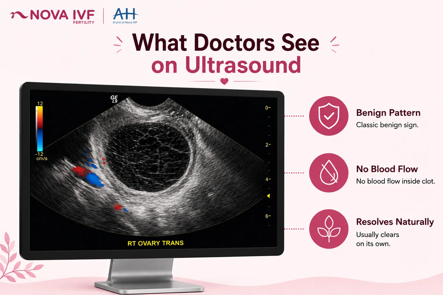
On a pelvic scan, a hemorrhagic cyst has a recognisable appearance. As the blood clot inside breaks down, fine strands of clotting protein form a lace-like, or reticular, pattern. The ACR O-RADS guideline, published in Radiology in 2020, records this reticular pattern and a retracting clot as classic benign features.
Common female fertility tests often include a transvaginal ultrasound, a scan performed with an internal probe that gives a clear, detailed view of the ovary. On colour Doppler, a setting that maps blood flow, the clot shows no internal flow. This absence of flow separates it from solid growth.
Hemorrhagic Follicle and Pregnancy: What You Need to Know
For most women, a hemorrhagic follicle is not associated with a lower chance of natural conception. These cysts are functional and usually clear within one or two menstrual cycles. Unlike an endometrioma, a functional hemorrhagic cyst leaves no lasting damage to ovarian tissue.
The term hemorrhagic follicle can be misleading. Bleeding most often occurs in the corpus luteum, the structure that forms only after the follicle has released its egg. A hemorrhagic corpus luteum therefore usually indicates that ovulation has happened, rather than that it has failed.
The corpus luteum also produces progesterone, the hormone that supports the lining of the uterus in early pregnancy. This progesterone output lasts about two weeks, extending only if an early pregnancy takes over. Bleeding into the gland does not stop its progesterone supply, so fertility is rarely affected.
During fertility treatment, a hemorrhagic follicle seen on a monitoring scan can affect the timing of a cycle. Time-lapse embryo monitoring and follicle tracking help the specialist plan around it. Patients should report any such finding so the cycle can be timed correctly.
Treatment and When to See a Doctor
Most hemorrhagic follicles need no active treatment beyond watchful waiting. A repeat ultrasound confirms that the cyst has shrunk or cleared. Pain is often mild and settles on its own; where relief is needed, a clinician can advise on suitable pain relief.
The ACR O-RADS guideline advises no routine follow-up for a typical hemorrhagic cyst up to 5 cm in premenopausal women. Cysts larger than 5 cm are usually rechecked with a scan after 8 to 12 weeks. In postmenopausal women, any hemorrhagic-looking cyst is less expected and is reviewed more carefully.
A few situations, though, are worth a same-day check with a doctor:
- Severe pain on one side that does not settle after a day or two warrants prompt review.
- Pain with dizziness, fainting, or a racing heartbeat can signal a ruptured cyst bleeding internally.
- A cyst that keeps growing, lasts beyond two cycles, or measures more than 5 cm needs assessment.
- Any new pelvic symptom during fertility treatment should be reported to the treating specialist.
Surgery, usually keyhole surgery called laparoscopy, is reserved for large, persistent, or ruptured cysts. A hemorrhagic follicle does not lower egg supply, unlike the challenges in a Nova case study, the Ovarian Reserve Journey.
Frequently Asked Questions
Is a hemorrhagic follicle the same as an ovarian cyst?
A hemorrhagic follicle is one type of ovarian cyst, a functional cyst that has filled with blood. It differs from cysts caused by conditions such as endometriosis, and from true tumours. Functional cysts like this usually clear on their own within a cycle or two.
Can a hemorrhagic follicle disappear on its own?
Yes, and most do. These cysts are part of the normal ovulatory cycle and usually clear within one or two months.
What does a hemorrhagic follicle look like on ultrasound?
It looks like a cyst with fine internal threads forming a lace-like or fishnet pattern as the clot breaks down. This reticular pattern is a classic benign feature [ACR O-RADS, 2020]. A transvaginal scan gives the clearest picture, and the clot shows no blood flow on Doppler.
Does a hemorrhagic follicle mean I did not ovulate?
Usually the opposite. A hemorrhagic corpus luteum forms from the structure left after an egg is released, so it generally means ovulation did happen. Persistent ovulation disorders, such as those seen in PCOS, are assessed separately.
Related Fertility Blogs