Self-cycleTM IVF

-
-
-
 Infertility Counselling
-
 Female Infertility Treatment
-
 Andrology Treatment
-
 Fertility Enhancing Surgeries - Female
-
 Fertility Enhancing Surgeries - Male
-
 Endoscopy Treatment
-
 IUI Treatment
-
 IVF Treatment
-
 ICSI Treatment
-
 Advance IVF Solutions
-
 Embryology
-
 Vitrification Egg, Embryo, Sperm Freezing
-
 Preimplantation Genetic Testing (PGT)
-
 Donation Program Embryo / Egg / Sperm
-
 Self-cycleTM IVF
-
-
-
-
-
Identifying Fibroids or Polyps: What You Should Know
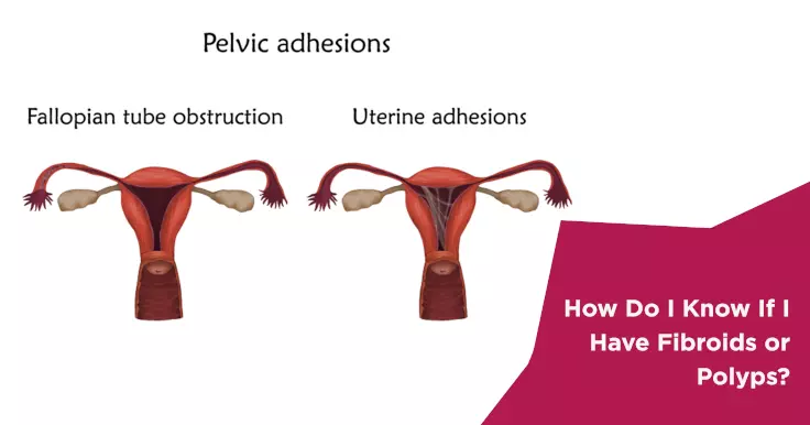
The uterus is made of many layers of muscles and tissues. Sometimes the cells in the muscles may begin to grow rapidly in one or more spots. This growth is termed as a fibroid. In other cases, the tissue cells of the innermost layer of the uterus or the endometrial layer may begin to overgrow in spots. This is referred to as a polyp. Fibroids and polyps can develop anywhere inside the uterus. These growths are usually benign and hence not something to worry about. In many cases, they may shrink on their own.
When is Treatment Needed?
Polys and fibroids can cause discomfort during menstruation. Painful abdominal cramps and heavy bleeding is usually a symptom of either of these growths. In the case of polyps, it may also cause spotting between periods. Treatment is usually sought only if these symptoms are very severe. This treatment usually takes the form of medication to relieve the symptoms. It is important to know that these medicines provide temporary relief and the symptoms may recur once the treatment is stopped.
Fibroids and polyps can develop anywhere inside the uterus.
Large fibroids can interfere with a woman's fertility. Thus, if a woman is finding it difficult to conceive and has fibroids, surgical treatment may be required to remove the fibroid completely.
Diagnosis of Uterine Fibroids and Polyps
A thorough physical examination of the pelvic area is the first step towards diagnosing fibroids or polyps. This may be followed by a few tests:
Ultrasound - Fibroids are usually visible in an abdominal ultrasound. This test uses sound waves to create an image of the uterus on a computer screen. The doctor may then identify fibroids and measure them to ascertain whether treatment is needed.
MRI - An MRI may be used to determine the location and size of fibroids.
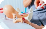
Vaginal Ultrasound - This involves inserting a flexible, wand-like device into the vagina. This device uses sound waves to create an image of the uterus on a screen. Any polyps or fibroids that may be present should be clearly visible in this image.
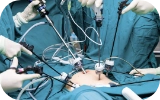
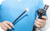
Hysteroscopy - This involves inserting a hysteroscope through the vagina into the uterus to give the doctor a clear view of the insides of the uterus. A hysteroscope can be described as a flexible, lighted telescope.
Hysterosonography - If the images created by an ultrasound are not clear, the doctor may advise a hysterosonography. This is also known as a saline infusion sonogram as it involves injecting a sterile saline solution into the uterus to expand the uterine cavity.