
-
-
-
 Infertility Counselling
-
 Female Infertility Treatment
-
 Andrology Treatment
-
 Fertility Enhancing Surgeries - Female
-
 Fertility Enhancing Surgeries - Male
-
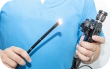 Endoscopy Treatment
-
 IUI Treatment
-
 IVF Treatment
-
 ICSI Treatment
-
 Advance IVF Solutions
-
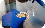 Embryology
-
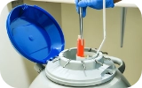 Vitrification Egg, Embryo, Sperm Freezing
-
 Preimplantation Genetic Testing (PGT)
-
 Donation Program Embryo / Egg / Sperm
-
-
-
-
-
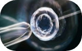
How Is Teratospermia Diagnosed? Explore Tests for Abnormal Sperm Shape

When a couple has been unable to conceive for over a year, visiting a fertility centre becomes essential. At the centre, the fertility parameters of both man and woman will be evaluated in order to diagnose the problem.
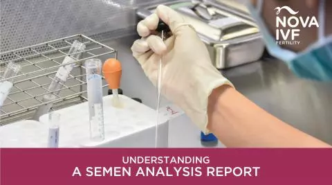
The standard examination for men is the seminogram, which is a comprehensive sperm analysis. This analysis is drawn from the semen sample provided by the man, and scrutinised under a microscope to detect anomalies, if any.
In order to diagnose teratospermia, a semen morphology analysis is conducted.
Diagnosis of Teratospermia
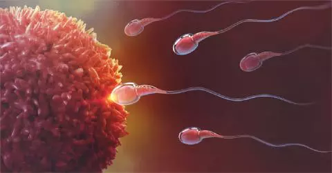
A normal shaped sperm has an oval head, a well-defined midpiece and a long tail. The World Health Organization [WHO] has issued strict criteria to evaluate teratospermia. Each part is investigated as follows:
- Head: The head has to be oval shaped and distinctly outlined. The acromosome and the nucleus have to be clearly visible as two parts of the head. There have to be less than 20% vacuoles.
- Midpiece: The midpiece has to be lineal. It is 1.5 time longer than the head. It is expected to have an equal proportion of thickness between the head and the tail.
- Tail: The tail has to be the slimmest part of the sperm, and only one in number.
It is important to note that even the ejaculation of a healthy, fertile male has as much as 50% of the sperm abnormally shaped. Therefore, there needs to be a really high proportion of teratospermia for it to affect the chances of conceiving.
WHO has also stated that teratospermia can be diagnosed if only 4% of the sperms show a normal shape.
Kruger's criteria, on the other hand, states that 15% of sperms should be normally shaped.
According to this criterion, diagnosis of teratospermia can be classified as the following:
- Mild teratospermia: number of normal shaped sperms ranges from 10% to 14%
- Moderate teratospermia: 5-9% are normal
- Severe teratospermia: Less than 5% have an ideal morphology.