 Self-cycleTM IVF


-
-
-
 Infertility Counselling
-
 Female Infertility Treatment
-
 Andrology Treatment
-
 Fertility Enhancing Surgeries - Female
-
 Fertility Enhancing Surgeries - Male
-
 Endoscopy Treatment
-
 IUI Treatment
-
 IVF Treatment
-
 ICSI Treatment
-
 Advance IVF Solutions
-
 Embryology
-
 Vitrification Egg, Embryo, Sperm Freezing
-
 Preimplantation Genetic Testing (PGT)
-
 Donation Program Embryo / Egg / Sperm
-
 Self-cycleTM IVF
-
-
-
-
-
Placenta Accreta: Meaning, Types, Causes and Treatment
Placenta Accreta: Meaning, Types, Causes and Treatment
Finding out that your pregnancy carries a risk of placenta accreta can feel overwhelming, but it is a manageable and well-understood high-risk condition when diagnosed early.
Placenta accreta is a pregnancy condition where the placenta attaches too deeply to the uterine wall. Doctors use "placenta accreta spectrum" as the umbrella term to describe how deep this attachment goes.
With timely prenatal planning and specialized care, healthcare teams can safely manage delivery for both mother and baby.
This guide covers the meaning, types, causes, symptoms, diagnosis, and modern treatment options for placenta accreta.
What Is Placenta Accreta? (Meaning and Overview)
Placenta accreta means the placenta has grown into the uterine wall more firmly than it should. In a normal pregnancy, the placenta lifts away cleanly after birth from a soft separating layer. With accreta, that layer is missing or damaged, so the placenta stays stuck.
Because it will not detach on its own, trying to remove it can cause heavy bleeding. In medical terminology, the same condition is sometimes called a morbidly adherent placenta. Both terms describe the same problem of abnormal uterine wall attachment.
The condition has grown more common in recent decades, largely because cesarean rates have risen. Clinical reviews now put its frequency at roughly 1 in 500 births. That rising trend is a large part of why awareness of it matters.
Placenta Accreta Spectrum: Accreta, Increta and Percreta
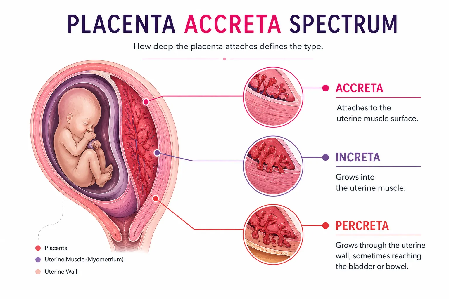
The placenta accreta spectrum is categorized into three distinct types of placenta accreta based on how deeply the placental tissue invades the uterine wall:
| Type | How deep it goes | Roughly how common | Main concern |
|---|---|---|---|
| Accreta | Attaches to the uterine muscle surface | About 75 to 80% of cases | Bleeding when the placenta will not separate |
| Increta | Grows into the uterine muscle | About 15 to 18% | Heavier bleeding, more complex surgery |
| Percreta | Passes through the wall, sometimes reaching the bladder or bowel | About 5 to 7% | Highest bleeding and organ-injury risk |
Each type of placenta accreta carries a different bleeding risk and level of surgical complexity. Accurate pre-delivery diagnosis helps maternal-fetal medicine specialists assemble the right surgical team, plan the delivery timing, and secure blood products well in advance.
Causes of Placenta Accreta and Key Risk Factors
The causes of placenta accreta usually trace back to a weak spot in the uterine lining. A scar from a cesarean or any other uterine surgery as for fibroids can leave exactly the kind of spot a placenta anchors into. The Society for Maternal-Fetal Medicine lists the main risk factors.
- A previous cesarean delivery, with the risk rising after each one
- Placenta previa, where the placenta extends over the cervix
- Earlier uterine surgery, such as fibroid removal or a D&C
- Maternal age above 35
- Several previous pregnancies
The highest single risk is combination of placenta praevia with a previous caesarean scar on the uterus. A 2006 study by Silver and colleagues in Obstetrics and Gynecology found that the risk on placenta accreta was about 3% after one caesarean and roughly 40% after three.
Pregnancies conceived through IVF also carry a higher observed risk of placenta accreta spectrum. A 2021 systematic review and meta analysis in institutional reports found that IVF embryo transfer was associated with approximately 5 times the odds of placenta accreta spectrum compared with spontaneous conception.
Symptoms of Placenta Accreta, Diagnosis and When to See a Doctor
Most cases of placenta accreta cause no symptoms and are first suspected on a routine scan. When a symptom does appear, the most common is painless vaginal bleeding in the third trimester, while pelvic pain is far less common.
Because the signs can be absent or subtle, any unexplained bleeding in pregnancy should be treated as urgent. Early contact with the medical team should be done to check the placenta without delay. Reach your maternity team straight away if you notice:
- Any vaginal bleeding in the second or third trimester
- Bleeding with or without pain
Diagnosis rests mainly on a detailed prenatal ultrasound, often with colour Doppler. It can show abnormal blood-vessel patterns and loss of the normal clear line between placenta and uterine muscle. These findings point to the condition before birth.
An MRI is advised when the ultrasound picture is unclear, or when percreta reaching the bladder is suspected. Even so, some milder cases are only confirmed at delivery. This is why history and imaging are always considered together.
In women with a previous caesarean and a low-lying placenta, the team looks closely for these signs from the mid-pregnancy scan on. A uterine fibroid or history of other prior uterine surgery raises the same alertness. Spotting it early converts a possible emergency into a scheduled, prepared birth.
Placenta Accreta Treatment and Delivery Planning
Placenta accreta treatment centres on a carefully planned birth rather than an emergency one. According to ACOG and the Society for Maternal-Fetal Medicine, the safest outcomes come from planning ahead at a specialist centre.
Note: This content is for informational purposes and does not substitute medical advice, since every case of pregnancy is different.
A well-prepared delivery plan usually includes:
- A scheduled cesarean, commonly observed between 34 and 37 weeks, before labour is likely to begin
- Delivery at a specialist centre with the right facilities and expertise
- A multidisciplinary team, including an obstetrician, a maternal-fetal medicine specialist, and surgical support
- Blood products kept ready in case heavy bleeding occurs
The 34-to-37-week window during pregnancy balances two risks. Delivering too early strains the baby, while waiting too long risks bleeding if labour starts on its own. Planning care at this level has been shown to lower the risk of severe bleeding and other complications.
A common misconception is that a hysterectomy (removal of the uterus) is necessary with every accreta diagnosis. While a cesarean hysterectomy is often the safest choice in extensive increta or percreta cases to prevent life-threatening hemorrhage, it is not the only outcome.
Small, localized (focal) accreta cases can sometimes be managed with uterine-preserving techniques, such as surgical oversewing of the attachment site or localized resection or by uterine artery embolization. Management decisions are made on a case-by-case basis, balancing patient safety, bleeding severity, and future fertility desires.
Complications of Placenta Accreta and Future Pregnancy
The main complications of placenta accreta come from bleeding when the placenta will not detach. Severe cases can involve large blood loss and injury to nearby organs. The planned early delivery also means many babies arrive a few weeks preterm.
The risks the team plans around include:
- Heavy bleeding that may need a blood transfusion
- Injury to the bladder or bowel in deep percreta
- Preterm birth from the planned early delivery
- A stay in intensive care after complex surgery
- Sometimes, severe bleeding may necessitate removal of uterus at the time of caesarean delivery (caesarean hysterectomy) in order to save the life of the mother
Listing these is not meant to alarm. Each of these complications of placenta accreta is exactly what a specialist team plans around, and good planning is what keeps them uncommon. The whole point of early diagnosis is to shrink this list before the birth ever happens.
Future pregnancy is still possible when the uterus is preserved. It does carry a higher chance of recurrence,, so it needs close monitoring from the very start. When a hysterectomy is needed, it ends the ability to carry a future pregnancy. However, the couple still carries the ability of having their own children with the help of surrogacy.
Anyone with risk factors, such as a previous cesarean or an IVF pregnancy, benefits from raising this early with a maternal-fetal medicine or fertility specialist. For fertility patients, the IVF pregnancy monitoring process already provides this kind of close, early oversight.
An early, personalised monitoring plan is the most useful step of all. Placenta accreta is far safer when it is expected than when it is found by surprise. Raising it early costs nothing and can change the entire course of the birth.
Frequently Asked Questions
What is the main cause of placenta accreta?
The most common cause is a previous cesarean scar. Damaged uterine lining lets the placenta attach too deeply, and the risk rises with each prior cesarean.
How serious is placenta accreta?
It is a high-risk condition because of the increased chances of bleeding involved. But when it is diagnosed early and delivery is planned by a specialist team, outcomes are much safer.
What is the difference between placenta accreta, increta and percreta?
They differ by depth. Accreta attaches to the uterine muscle, increta grows into it, and percreta passes through the muscular wall, sometimes reaching nearby organs.
Can placenta accreta be detected before delivery?
Yes. A detailed prenatal ultrasound detects most cases, with MRI used for deeper or unclear ones, letting the team plan a safe delivery in advance.