 Self-cycleTM IVF

-
-
-
 Infertility Counselling
-
 Female Infertility Treatment
-
 Andrology Treatment
-
 Fertility Enhancing Surgeries - Female
-
 Fertility Enhancing Surgeries - Male
-
 Endoscopy Treatment
-
 IUI Treatment
-
 IVF Treatment
-
 ICSI Treatment
-
 Advance IVF Solutions
-
 Embryology
-
 Vitrification Egg, Embryo, Sperm Freezing
-
 Preimplantation Genetic Testing (PGT)
-
 Donation Program Embryo / Egg / Sperm
-
 Self-cycleTM IVF
-
-
-
-
-
Everything You Need to Know About Scrotal Ultrasound
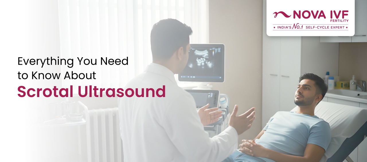
The scrotal ultrasound is one of the primary methods of evaluating any disorders of the testicles, the epididymis, and other scrotal structures. Generally recommended by an andrologist, a scrotal ultrasound can be helpful in determining the cause of infertility in males as well. It is a fairly simple procedure, but it can reveal valuable information about a male’s reproductive health. Naturally, a person who has been recommended to get a scrotal ultrasound may feel curious to know about the procedure. Here, you can learn everything you need to know about scrotal ultrasound and what to expect during this test.
Synopsis
A scrotal ultrasound is a simple and painless testicular ultrasound used to check the testicles, epididymis, and surrounding scrotal structures. It helps doctors detect problems like swelling, infection, cysts, torsion, and varicocele diagnosis. It is also commonly advised as an ultrasound for male infertility evaluation. In this blog, you will learn what is a scrotal ultrasound, why it is done, and during a scrotal ultrasound, what to expect.
What Is a Scrotal Ultrasound?
Scrotal ultrasound, also known as testicular ultrasound, is a diagnostic imaging test that is used to obtain images of the testicles and surrounding tissue. This is a non-invasive test that can help your andrologist diagnose medical conditions affecting the organs contained in the scrotum.
The scrotum is a skin-covered, muscular sac, and it is located at the base of the penis. Scrotum is essentially a sac that holds the testicles of a male. Testicles are an important part of the male reproductive system that are responsible for producing testosterone (one of the male sex hormones) and sperm. The scrotum contains epididymis, blood vessels, and vas deferens (a coiled tube that carries the sperm out of the testes).
Testicular ultrasound is used to examine the testicles and other scrotal structures. The results of the ultrasound procedure are visible on a monitor, which aids the technician in properly examine the scrotal structures. Oftentimes, scrotal ultrasound is used to determine the cause of enlargement of the testicles or pain in the testicles.
The testicular ultrasound is a safe, painless procedure that captures the inside of the scrotum using sound waves. High-frequency sound waves travel through the gel into the body from the small probe.
Purpose of the Scrotal Ultrasound
The scrotal ultrasound is typically used to:
- Diagnose the cause of scrotal pain that is commonly caused by epididymitis.
- Check for absent or undescended testicles.
- Check for twisting of the spermatic cord or testicular torsion that can be caused by some abnormalities during foetal development.
- Determine the location and condition of the mass or lump found in the scrotum during a physical examination
- Evaluate the reason for infertility that may be caused by conditions such as varicocele
- Measure the testicular size
- Check the testicles of men with a history of testicular cancers or infection
- Serve as a guide during needle biopsy for certain testicular masses
Why Do I Need a Scrotal Ultrasound?
Your doctor will recommend a scrotal ultrasound if you are experiencing any pain, swelling, or other concerning symptoms related to your scrotum. The scrotal ultrasound can help diagnose the following health problems:
1. Testicular Torsion
If there is a sudden onset of pain in a sensitive area like the scrotum, it is usually a cause for concern and shouldn’t be ignored. Testicular torsion is a condition in which the spermatic cord, which supplies blood to the testicle) becomes twisted and leads to sudden and severe pain in one testicle.
This is a serious medical condition that required immediate medical attention to avoid the death of the tissue of the affected testicle as it is not receive any blood supply. Surgery is often required to treat the condition and prevent further damage to the testicle.
2. Testicular Lumps
A testicular lump revealed in a physical examination may be a cancerous tumour. The doctors recommend a scrotal ultrasound if they suspect any masses or lumps inside the scrotum. The result of a scrotal ultrasound can help the doctor evaluate the lump for its size, location, and other parameters. If the lump appears to be solid in the ultrasound images, it can be cancerous, but if the lump is filled with fluid, it is most likely harmless.
3. Epididymitis
Epididymitis (inflammation of the epididymis) can be the cause of pain in your scrotum as it is one of the most common causes of pain in that area. Epididymitis can lead to fluid build-up around the testicle and cause swelling or lump formation. However, this condition is easily treatable with the help of antibiotics as it is often caused by an infection.
4. Infertility
A scrotal ultrasound may also be a part of an infertility assessment that you may require in order to proceed with your fertility treatment. Since the testicles are responsible for making and storing sperm, any problem affecting this area can cause infertility. Thus, it is important to examine the area properly to understand the cause of infertility for a male.
5. Undescended Testicles
Scrotal ultrasound is also an excellent tool to detect an absent or undescended testicle. An undescended testicle is common in young males. When a male foetus is developing inside the womb, his testicles ideally move downwards to sit outside the body below his penis from the abdomen. This process usually happens before the baby is delivered, but it may take up to 6 months after birth to happen for a male infant.
In case the baby boy’s testicles don’t descend on their own within 6 months of his life, it is essential to consult a specialist. The doctor will likely recommend a scrotal ultrasound to detect the presence and location of the undescended testicles. To treat undescended testicles, surgery may be required to manually move the testicles down to sit in the correct position.
FAQs
1. What is a scrotal ultrasound?
A scrotal ultrasound is a scrotal imaging test that uses sound waves to create images of the testicles and nearby structures. It is also called a testicular ultrasound.
2. For a scrotal ultrasound, what to expect during the test?
During the scrotal ultrasound procedure, gel is applied and a probe is moved over the scrotum. The test is usually painless, but mild discomfort may occur if the area is tender.
3. What does a scrotal ultrasound show?
It can show testicular swelling, fluid collection, cysts, infection, blood flow issues, and abnormal masses. It is also useful for varicocele diagnosis.
4. What is a scrotal ultrasound used for in infertility?
In many cases, a scrotal ultrasound is advised as an ultrasound for male infertility. It helps doctors look for problems such as enlarged veins (varicocele), reduced testicular size, or other scrotal findings that may explain a low sperm count.
5. Is scrotal ultrasound safe?
Yes. A scrotal ultrasound is a safe scrotal imaging test. It does not use radiation and is generally painless and without side effects.
6. Do I need any preparation before a scrotal ultrasound?
Usually, no. For the scrotal ultrasound procedure, you are only asked to change into a gown and lie down comfortably while the scan is performed.