


-
-
-
 Infertility Counselling
-
 Female Infertility Treatment
-
 Andrology Treatment
-
 Fertility Enhancing Surgeries - Female
-
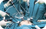 Fertility Enhancing Surgeries - Male
-
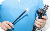 Endoscopy Treatment
-
 IUI Treatment
-
 IVF Treatment
-
 ICSI Treatment
-
 Advance IVF Solutions
-
 Embryology
-
 Vitrification Egg, Embryo, Sperm Freezing
-
 Preimplantation Genetic Testing (PGT)
-
 Donation Program Embryo / Egg / Sperm
-
-
-
-
-
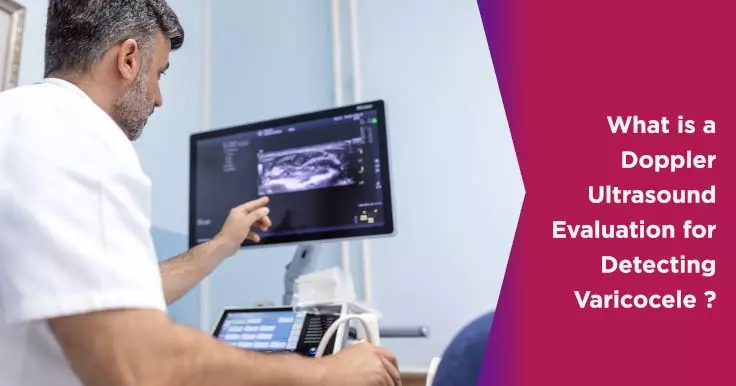
Varicocele Grades and Doppler Ultrasound Evaluation: A Comprehensive Guide
The testicles are contained in a loose bag of skin. Enlargement in the veins in this part of the skin is known as a varicocele. This can be compared to varicose veins that affect the legs. In some cases, varicoceles can cause blood to flow backward and hence lower sperm quality and reduce sperm production causing infertility. They can also make the testicles shrink or keep them from developing normally. In such cases, surgery may be needed to fix the enlarged vein. Thankfully, a varicose vein is fairly easy to diagnose and cure.
Doppler Ultrasound Evaluation
When diagnosing a varicocele, the doctor will usually follow a physical examination with an ultrasound. This is mainly done in cases where a physical examination is inconclusive. The Doppler ultrasound uses high-frequency sound waves to create images of the internal organs on a computer screen. This allows the doctor to identify problem areas without performing open surgery. It also helps doctors rule out other causes such as tumours for the symptoms being experienced.
This type of varicocele test can be used to detect nearly all varicocele cases. The dilated veins can be seen and measured on the images generated by the ultrasound. In mild cases, the diameter of the veins may not be enough to diagnose the condition but may be used to document the pathology. In cases of advanced varicocele, the diameter of the affected veins can range from 2-3 mm.
Varicocele tests can also be used to assess the hemodynamic changes associated with this condition. This can help confirm a diagnosis especially in cases where the diameter of the vein is quite small. A Valsalva manoeuvre may be performed to see this change. This involves standing upright and breathing out while closing the mouth and pinching the nose. The closed airways increase intra-abdominal pressure and thus make blood flow patterns more clearly visible.
There are many different patterns that have been identified in the blood flow of these dilated veins. The most specific amongst them is a venous flow wherein the blood flows towards the testis and increases abdominal pressure.
Varicocele Grading
There are 5 grades of varicoceles that may be categorized based on a Doppler ultrasound.
- Grade I - In such cases, there is no visible dilation in the intrascrotal veins but a reflux may be seen in the spermatic cord veins while performing a Valsalva manoeuvre.
- Grade II - The veins at the upper pole of the testis appear prominent and a reflux may be seen in the upper pole veins during a Valsalva manoeuvre.
- Grade III - While the patient is lying down, no major dilation may be noted but in a standing position the veins of the lower pole of the testis may appear dilated. A reflux may also be noted at the lower pole of the testis during a Valsalva manoeuvre.
- Grade IV - In such cases, the veins appear dilated even when the patient is lying down. A reflux may also be noted at the lower pole of the testis during a Valsalva manoeuvre.
- Grade V - Varicocele in this grade appears dilated when the patient is lying down as well as when standing and a reflux can be seen even without the Valsalva manoeuvre.
Related Fertility Blogs